Implantable computers
Chris Diorio
Neurons and neuronal networks decide,
remember, modulate, and control an animals every sensation, thought, movement,
and act. The intimate details of this network, including the dynamical
properties of individual and populations of neurons, give a nervous system the
power to control a wide array of behavioral functions; the unknown character of
much of this detail motivates many workers in modern neurobiology.
Neurobiologists examine the activities of brain cells tied to sensory inputs,
integrative processes, and motor outputs, to understand the neural bases of
behavior. They also probe the components of neuronal control circuitry, to
understand the plasticity and dynamics of control. They want to know more about
neuronal dynamics and networks; about synaptic interactions between neurons;
about how neuronal signaling and behavior and control and environmental stimuli
are inextricably linked.
To make significant progress,
neurobiologists need methods for recording the activity of single neurons or
assemblies of neurons, for long timescales, at high fidelity, in animals that
are free to interact with their sensory world, and that are free to express
normal behavioral responses. Contemporary tools for studying neuronal signaling
and information processing include imaging at the cellular and anatomical levels
(e.g. single-photon microscopy, voltage-sensitive dyes, PET scans, etc.), direct
electrical measurement using micropipettes or micromachined extracellular
probes, and a host of other techniques. But the one tool that can truly help
unravel the neural substrates of behaviorthe digital computeris, at present,
utterly dissociated from nerve tissue. Neurobiologists use computers for
simulation and for data collection, but not to probe the neurobiology
directlythat is, they have not linked the electronic signaling of digital
computers directly with the electronic signaling of nerve tissue.
Our research focuses on direct electrical
measurement (and stimulation) of nerve tissue, primarily because voltage
signaling is the lingua franca of both nerve tissue and computersboth use
time-varying voltage waveforms to convey and to process information.
Neurobiologists employ two classes of electrodes to record and stimulate
electrical signals in tissue: (1) intracellular micropipettes for single-site
intracellular and patch-clamp interrogation of neurons (one site per pipette),
and (2) extracellular wires or micromachined probes, for interrogating multisite
patterns of extracellular neural signaling or electrical activity in muscles.
For recording in neurons, intracellular probes afford roughly a thousand-fold
larger signal-to-noise ratio than do extracellular probes. However, multi-site
recording is key to gaining access to patterns of neuronal activity. Currently,
high-fidelity recording of neuronal signals is practical only on small numbers
of neurons, in constrained laboratory experiments using immobilized animals,
primarily because there are no probes for multi-site intracellular recording.
For recording in muscles, extracellular probes allows adequate signal fidelity;
the goal is to record or stimulate during normal behavior, so as to investigate
the neuromechanical systems that underlie the behavior.
Tom Daniel ,
Denice Denton
,
Karl Böhringer
,
Dennis Willows
, and I
have initiated a multi-disciplinary research program
to take the first steps toward integrating computer
electronics with neurobiology. More specifically, we are (1) implanting a
standalone microcomputer into the brain of a marine mollusc, to allow multi-site
intracellular recording and stimulation in a live, freely behaving animal, and
(2) embedding a standalone microcomputer into the sensory and muscle/control
pathways of a giant moth, to probe the dynamical control of flight in a live,
freely behaving animal. The compelling scientific reason for this research is to
correlate neuronal signaling and control with environmental stimuli and
behavior, to better understand the neural substrates of behavior.
Our research comprises four main thrusts:
(1) developing a standalone implantable microcomputer that records from and
stimulates multiple neurons or muscle fibers in an intact animal, using
intracellular probes, extracellular probes, or wire electrodes; (2)
developing micro-electromechanical (MEMS)
probe arrays for recording intracellularly from multiple neurons simultaneously
; (3) developing neurophysiological preparations and
techniques for implanting microchips and wire electrodes or MEMS probes into or
onto animals without damaging probes or tissue, including dealing with issues of
biocompatability, and (4) developing analytical models of the biology, and
experiments that software-test these models in behaving animals.
Our research focuses on Manduca sexta and
Tritonia diomedea, for several reasons. Both presently serve as model systems
for probing the neural bases of behavior. Both afford accessible recording
sites, with high signal fidelity. Manduca's flight circuits are relatively well
understood in the context of constrained laboratory environments. Tritonia has
extraordinarily large brain nerve cells, identifiable sensory and motor
functions associated with these brain cells, and robust response to surgical
insult. And, appearances to the contrary, the microcomputers that we are
developing will be compatible with both animals.
A. Manduca Sexta
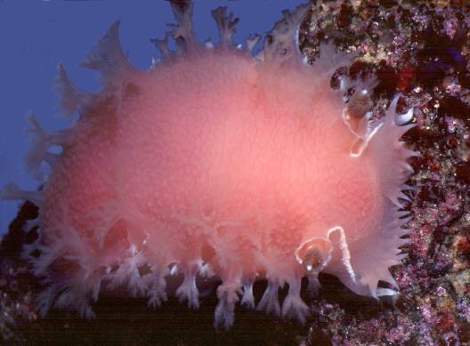
B. Tritonia Diomedea
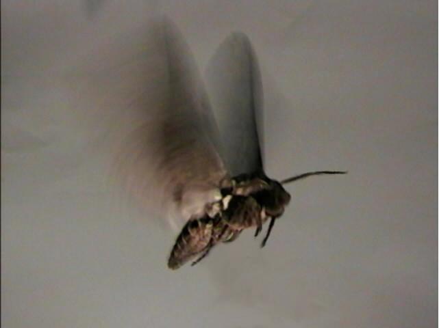
Manduca Sexta is typically 4cm in length, with a 12cm
wingspan; at 2.5g, it is among the largest of insect flyers. It can easily
carry a test-electronics payload. Tritonia Diomedea is typically 20cm in
length, and has a readily accessible brain with large and well-characterized
neurons.
back
to Chris Diorio's home page